The team started work on the project in March 2020, just as the pandemic hit. Working from home with CT scan images of healthy dogs supplied by the Vet School’s image archive, Richard started making prototypes on his home 3D printer.
The project team met on Zoom and Teams, refining the prototypes over various iterations. When restrictions eased in late summer 2020, Richard was once again able to access ECA’s high-specification 3D printers to hone and perfect the model through experimentation and testing.
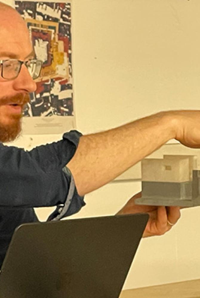
The result was a life-sized and anatomically precise 3D-model of a dog’s lumbar vertebrae – bones in the lower back – and pelvis.
This 3D-model was augmented with synthetic materials and a system of fluids which was placed inside a dummy dog that can be positioned in exactly the same way as patients undergoing this procedure.
Experts who took part in validating the model, which was also tested by vet students, reported that it was realistic and a good representation of a living patient.
There are many benefits to ECA students resulting from staff undertaking research of this type and collaborating with colleagues from across the University. Richard says, “It was a win-win-win situation: it was good for the Vet School, for our department at ECA, and for our students, who get the chance to see the process of making sophisticated and complex models. They are also benefiting from the knowledge I gained through this type of experimentation.”
Richard’s collaborators at the Vet School were Dr Megan Madden (Resident in Small Animal Neurology & Neurosurgery), Dr Tobias Schwarz (Reader in Diagnostic Imaging, DI Service Head), and Dr Anna Suñol (Senior Lecturer in Veterinary Neurology).
The team say their design is a useful proof of concept for the application of 3D printing in developing anatomical models for teaching purposes.
Their design is available to all, enabling future studies to develop or adapt it for similar or alternative procedures.
The study is published in the Journal of Veterinary Medical Education.